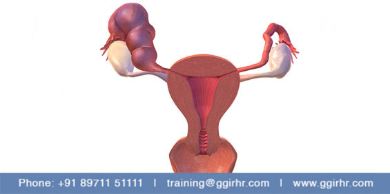
HYDROSALPHINX
A hydrosalpinx is a blocked, dilated, fluid-filled fallopian tube
Hydrosalpinx may occur as an isolated adnexal lesion or as one component of a complex adnexal lesion that has caused distal tubal occlusion . The most common cause of distal tubal occlusion and hydrosalpinx is pelvic inflammatory disease. Other causes include endometriosis, peritubal adhesions from a previous operation, tubal cancer, and tubal pregnancy.
Ultrasound
• thin- or thick-walled (in chronic cases)
• elongated or folded, tubular, C-shaped, or S-shaped fluid-filled structure
• distinct from the uterus and ovary.
.The folds may produce a characteristic “cogwheel” appearance when imaged in cross section. These folds are pathognomonic of a hydrosalpinx. Indentations on the opposite sides of the wall is referred to as the waist sign which is a strong predictor of hydrosalpinx. The waist sign in combination with a tubular-shaped cystic mass has been found to be pathognomonic of a hydrosalpinx .The ‘beads-on-a-string’ sign is described as hyperechoic mural nodules measuring about 2–3 mm and seen on cross-section of the fluid-filled distended structure
Sometimes the dilated fallopian tube may not show longitudinal folds. If the elongated nature of these folds is not noted, they may be mistaken for mural nodules of an ovarian cystic mass. A significantly scarred hydrosalpinx may present as a multilocular cystic mass with multiple septa (often incomplete) creating multiple compartments. These septa are generally incomplete, and the compartments can be connected. However, with more pronounced scarring, differentiation from an ovarian mass may not be possible.
(a) “Waist sign” of a hydrosalpinx, marked by the asterisks, as seen with 2-D ultrasound.
(b) “Beads on a string” sign of a hydrosalpinx demonstrated by 2-D ultrasound
(c) Sagittal view -nodular appearance of longitudinal folds at junction of collapsed and distended segments
(d)Transverse sonograms show distended funneled distal end of hydrosalpinx
(e) Image showing incomplete septae
(f) No flow seen with color Doppler
(g) Cogwheel sign-swollen walls and swollen mucosal folds
Numerous studies have shown that hydrosalpinges have a detrimental effect on IVF success rates. Two meta-analyses of these studies noted that the pregnancy, implantation, and delivery rates were approximately 50% lower and that the spontaneous abortion rate was higher in the presence of hydrosalpinges . This finding may be due to mechanical flushing of the embryos from the uterine cavity, decreased endometrial receptivity, or a direct embryotoxic effect . Patients with hydrosalpinges visible on ultrasound may be more significantly affected . Randomized clinical trials (RCTs) comparing pregnancy rates and outcomes with IVF for women with hydrosalpinges, with or without prior laparoscopic salpingectomy, reported that salpingectomy restores the rates of pregnancy and live birth to levels similar to those of women without hydrosalpinx
A Cochrane analysis concluded that laparoscopic salpingectomy or occlusion should be considered before IVF for women with communicating hydrosalpinges . Even patients with a unilateral hydrosalpinx have been shown to have lower pregnancy rates with IVF . Unilateral salpingectomy resulted in a significant improvement in IVF pregnancy rates in these patients . However, salpingectomies for bilateral hydrosalpinges yielded higher IVF pregnancy rates than for unilateral hydrosalpinges


