
Siora Surgicals Pvt. Ltd. is a manufacturer and exporter of orthopedic implants and instruments for over three decades. We have a comprehensive portfolio of trauma products numbering more than two thousand.
A simple radiolucent table or a fracture table can be used for the operative treatment. The patient can be positioned either supine or lateral. Selection of position depends on the preference and experience of the surgeon. For intramedullary nailing, the image intensifier in two planes will be required so that the proximal end of the femur can be accessed.
Manual traction or distraction can be used for attaining closed reduction of femoral shaft fractures using a fracture table or distractor. Depending on the level of the fracture, abduction, or adduction can be applied by using a distractor. For intramedullary nailing, a short nail can be used as a joystick to manage the proximal fragment. The Ligamentotaxis technique can be used for the reduction of complex fractures.
Approaches
For antegrade femoral nailing, generally, a longitudinal incision of nearly 3–5 cm placed 12–15 cm proximal to the tip of the greater trochanter is adequate enough. The certain entry point which changes according to the design of the Antegrade Interlocking Nail can be exposed through a blunt dissection down to the tip of the greater trochanter. Fluoroscopy can be used for conventional antegrade locked nailing of mid-third and distal diaphyseal fractures as it helps in alignment of the proximal starting point with the medullary canal of the femur in two planes.
In the case of open plating, the skin incision should be on the lateral side of the thigh between the greater trochanter and the lateral femoral condyle. Then fascia lata is split and the vast lateral muscle is retracted together with intermuscular septum down to the linea aspera protecting the perforating vessels. In case of less invasive exposures in plating, the entry point of the plate should be at the lateral femoral condyle, where an incision of 3–5 cm is made anterolaterally. After the indirect reduction (femoral distractor), the sub-muscular route including the shaft of the femur is prepared with an elevator for inserting the plate. Small separate incisions can be made for fixation screws.
Selection of Implant
The determining factors for the implant selection are: configuration and location of the fracture, condition of soft-tissues, patient’s condition, multi-trauma, injury severity score(ISS), experience and preference of the surgeon, size of the medullary canal, presence of other implants, intraoperative imaging and availability of implants, instruments.
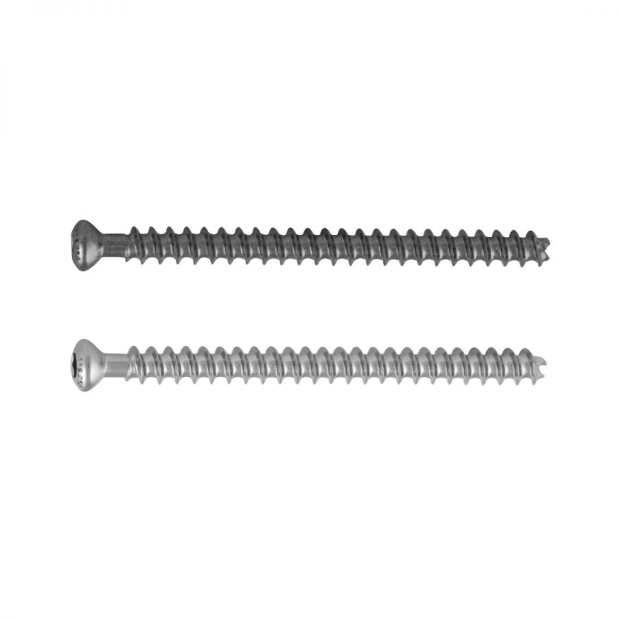
Generally, intramedullary nailing is used in case of diaphyseal fractures Condylar plates, dynamic condylar screws (DCS), solid femoral nails (UFN), and proximal femoral nails (PFN) can be used in case of subtrochanteric fractures. The universal or the new cannulated screws are used for fixing the simple fractures (type A and B) of the mid-third with interlocking technique and after reaming of the canal. Whereas stabilization of complex type C fractures and fractures of the proximal and distal third is achieved with the solid or cannulated intramedullary nail. In the rare situation of femur plating, the broad LC-DCP 4.5, the long condylar plates, or the dynamic condylar screw can be used.
Whereas external fixation or unreamed or minimally reamed intramedullary nailing are suggested for severe soft-tissue injury whether it is open or closed. External fixation is preferable for fixation of fracture in the severely multi-traumatized patient (with an ISS greater than 40) as least systemic and local interference is caused by external fixator. A more stable internal fixation device must be changed within 1–2 weeks else there may be the risk of pin-tract infection.


 4274
4274