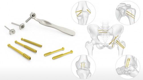
Siora Surgicals is India’s leading manufacturer and supplier of Orthopedic Implants. Siora Surgicals as Orthopedic Implants Manufacturer offers an exclusive range of trauma Implants and Instruments.
Plate fixation is suitable for tibia as the contouring of the plate is made easy by the flat medial surface. Additionally, when the fixation of plate is done along its medial subcutaneous surface, the plate does not cause any disruption in the critical blood supply to the bone. But plate contouring is challenging on the lateral surface. Although the lateral surface can be accessed yet separation of muscles is needed for the exposure and nerves and vessels have to be taken into consideration specifically.
Preoperative planning and approaches
The basic instrument set must include a range of narrow DCPs 4.5 or LC-DCPs 4.5, and reduction instruments for plating in the tibia. The patient should be in a supine position on a regular operating table (preferably a radiolucent one). An inflated cuff on the thigh is suggested. Generally, the tourniquet is not used. The narrow LC-DCP 4.5 and the PC-Fix are specially designed for the preservation of the periosteal blood supply. Moreover, due to their minimal contact with the bone, it lends itself to extra-periosteal positioning.
However, the standard approach to the tibia lies 1–2 cm lateral to the tibial crest but it may be medial in rare cases. An incision is made directly over the crest and ends up over the medial surface after skin closure and subsiding of the swelling due to the extra bulk of the plate. Though for the lateral approach, the skin incision to the tibia is similar to the medial side. The incision is made at the fascia overlying the muscle a few millimeters away from the crest so that fringe can be left for later reattachment. The muscles are separated gently from the tibia for positioning the plate.
Reduction techniques
The most significant part of the internal fixation is to select the correct technique for reduction. The goal of fixation must be to achieve proper alignment and rotation of the limb axis in all planes whether achieved by direct or indirect means. For the achievement of reduction, gentle and atraumatic manipulations must be used such that the critical blood supply to the fracture fragments is not compromised. The direct anatomical reduction should be followed in case of simple fracture patterns such as spiral, oblique, and bending or spiral wedges and plating should be done following the classical AO principles using interfragmentary cannulated lag screw fixation.
The exact reduction is not needed in complex fractures (Type C). The plate is required to only bridge the area of the fracture and minimal exposure and indirect reduction technique are followed. Restoration of adequate length, axial alignment, and rotation is imperative.
Choice of implant
The narrow DCP 4.5 or LC-DCP 4.5 is the implant of choice for diaphyseal fractures of the tibia. Minimum six cortices are needed on either side of the fracture in standard plating. Sometimes, smaller plates like DCP 3.5 are suggested in the distal tibia but these plates are not strong enough so should not be used as single implants. The rule of the six cortex is also applied in such cases. But broad plates are bulky and too stiff so they must not be used.
Nowadays longer plates with 8-10 holes are used (not to fill each hole). If screws are spaced apart and anchored in the bone of good quality, then 2-3 screws above and 2-3 screws below the fracture are considered sufficient. More screws should be used only if required.


