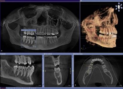
Today, Cone Beam Computed Tomography (CBCT) scanners are becoming a rage in dental imaging.
Today, Cone Beam Computed Tomography (CBCT) scanners are becoming a rage in dental imaging. It is used to plan dental implant treatment, endodontics, orthodontics, and maxillofacial surgery, etc. In general terms, the San Diego dentist uses it to scan the abnormal teeth and evaluate the jaws and facial structure. These scanners are a boon to the dentistry if used correctly. They can be are highly capable of being used in 3-D cone beam scanning techniques in coronal, sagittal, and axial planes. CBCT radiation exposure is ten times less in comparison to traditional CT scans.

How Do They Work?
CBCT scanner uses a fundamental principle of generating x-rays in a tube containing two charged electrodes opposite in nature (a cathode and an anode) separated by a vacuum in arranged in an electrical circuit. When the cathode is supplied with current, the filament heats up resulting in electron release. The high voltage is generated between electrodes causing the electron release to accelerate at high speed towards the anode colliding with each other at the focal point. The focal point in a CBCT scanner is generally 0.5 mm wide and determines the image's sharpness.
Does Patient Require 3-D Cone Beam CT Scan?
While radiation produced by CBCT scanners is minimal, it is recommended by the FDA to perform imaging modality only if it is clinically necessary. Here are some factors that San Diego dentist consider:
- Ensure that the information that is provided cannot be obtained by using other imaging modalities.
- All the benefits and even potential risks should deeply be analyzed before taking a CBCT scan in the patient case.
- Optimize the settings of exposure during the scan to provide the lowest radiation dose to give an adequate image quality.
This is a little snippet of a 3D cone beam scan explaining how it affects the modern dentistry system.


